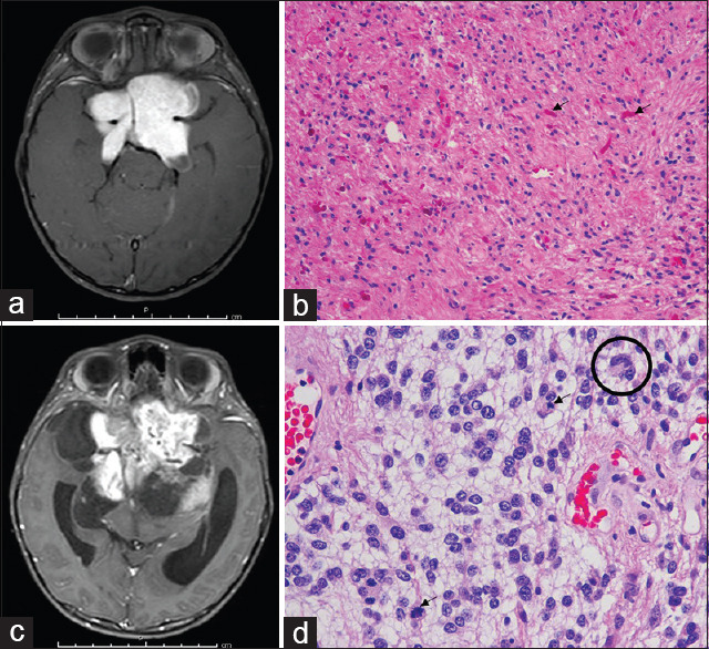
Figure 2.

Anaplastic transformation illustrative case of pilocytic astrocytoma. (a) Preoperative T1 weighted postcontrast magnetic resonance imaging showing an enhanced suprasellar mass. (b) H and E stain showing astrocytic cells neoplastic astrocytes in the glial fibrillary background, with numerous Rosenthal fibers (arrows). (c) T1 weighted postcontrast magnetic resonance imaging at 3 years later showing the larger residual tumor. (d) H and E stain showing an anaplastic transformation, including increased cellularity and pleomorphism of tumor cells with multinucleated cells (circle) and mitoses (arrows)
