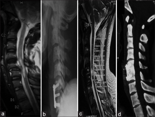
Figure 8.

(a) Magnetic resonance imaging T2-weighted sagittal images showing subluxation of the C5–C6 vertebrae causing compression of the cervical cord along with cord signal changes. (b-d) Postoperative sagittal X-ray, computed tomography, and magnetic resonance imaging T2-weighted images showing decompression and realignment of the cervical cord with fixation using cervical plates and screws
