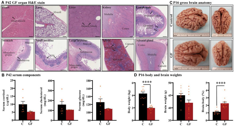
Figure 1.
General characterization of germ-free piglets. (A) H&E stains of germ-free (GF) organs at postnatal day P42. Scale bars, 500 μm. (B) Quantification of cortisol, cholesterol, and glucose levels in the serum of GF and control (C) animals at P42. n = 4–7 animals/group. (C) Gross anatomy of a GF and a control animal at P16. (D) Quantification of the total body weight, brain weight, and percent brain to body weight ratio at P16. Individual male and female animals are marked by orange triangles and circles, respectively. n = 11–12 animals/group. Data are expressed as mean ± SEM. ****p < 0.0001, unpaired student’s t-test for body weight and brain weight, Mann-Whitney U test for percent brain to body weight ratio.

