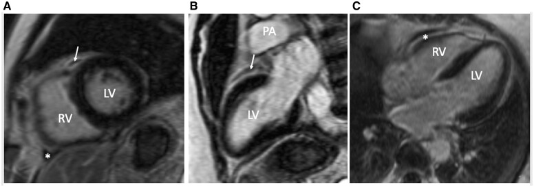
Figure 2.
Cardiac magnetic resonance imaging with late gadolinium enhancement: (A) cardiac short-axis view; (B) long-axis two-chamber view; (C) long-axis four-chamber view. A small local lens-like collection of pericardial fluid in front of right ventricular outflow tract is marked with arrows. Focal enhancement of adjacent pericardial layers is marked with an asterisk. LV, left ventricle; PA, pulmonary artery; RV, right ventricle; RVOT, right ventricle outflow tract.

