
Figure 6. Effects of IL‐6 on connexin43 protein expression in HL‐1 cardiomyocytes and Raw264.7 macrophages in culture.
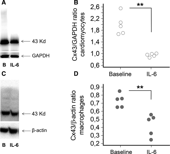
A, Western blot for connexin(Cx)43 in cardiomyocytes at baseline (B) and after treatment with IL‐6 (100 pg/mL) for 24 hours, and (B) the corresponding histograms for Cx43 normalized to GAPDH (n=5; mean percentage decrease=54%). Two‐tails Mann‐Whitney test: **P<0.01. C, Western blot for Cx43 in macrophages at baseline (B) and after treatment with IL‐6 (100 pg/mL) for 24 hours, and (D) the corresponding histograms for Cx43 normalized to β‐actin (n=5; mean percentage decrease= 43%). Two‐tails Unpaired “t” test: **P<0.01.