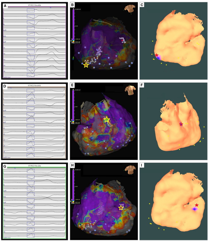
Figure 3. Localization of 3 premature ventricular contraction (PVC)‐origin sites by the automatic arrhythmia‐origin localization system.

A, D, G, Recorded 12‐lead ECG of PVCs during the procedure for a patient with arrhythmogenic right ventricular cardiomyopathy. B, E, H, Epicardial substrate map for this patient, with the PVC‐origin site (identified by activation and pace mapping) depicted by the yellow star. C, F, I, Usage of the automatic arrhythmia‐origin localization system to predsict a PVC origin site indicated by the blue patch onto the epicardial electroanatomic mapping surface, with the actual site of PVC origin marked by the red star.
