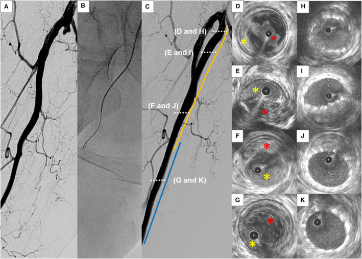
Figure 2. Representative case of subintimal approach.

A, Angiography shows a chronic total occlusion (CTO) lesion at the right femoropopliteal artery. B, Subintimal approach is attempted with a 0.035‐inch guidewire. C, Two bare‐metal nitinol stents (orange and blue lines) are successfully implanted. D through G, Intravascular ultrasound (IVUS) images of the wire passage within the CTO lesion from proximal (D) to distal (G). The guidewire is located in an intraplaque space (red asterisk) at the proximal portion of the lesion (D), whereas it goes through a subintimal space (yellow asterisk) thereafter. H through K, IVUS images after stent deployment from proximal (H) to distal (K).
