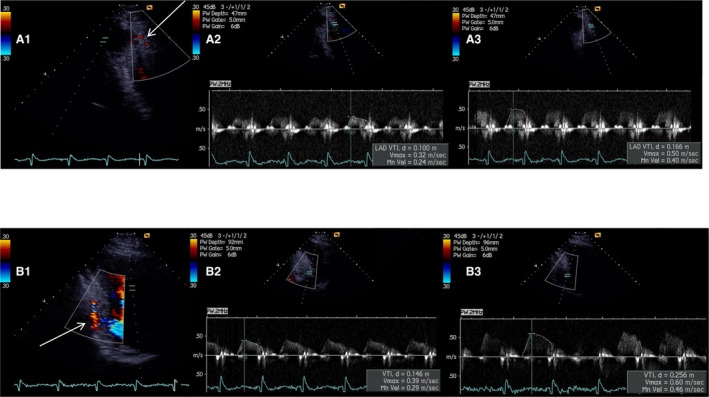
Figure 1. Examples of noninvasively derived coronary flow velocity reserve (CFVR) for left anterior descending artery (LAD) and posterior descending artery (PD).

(A1) Color Doppler signal of coronary flow for the distal segment of LAD in modified 3 chamber view (arrow). Peak baseline (A2) and hyperemic (A3) diastolic flow velocities with impaired CFVR LAD (0.50/0.32) – 1.56. (B1) Color Doppler signal of coronary flow for the PD, in modified 2 chamber view (arrow). Peak baseline (B2) and hyperemic (B3) diastolic flow velocities with impaired CFVR PD (0.60/0.39) – 1.54.
