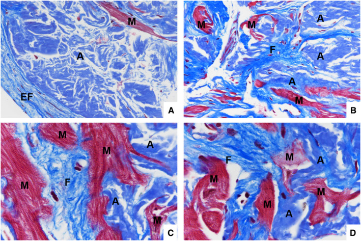
Figure 2. Distribution of amyloid deposits and fibrosis.

Endocardial (EF) and interstitial (F) fibrosis in the left ventricle endomyocardial biopsy from a transthyretin‐positive (A and C) and a lambda+ AL (B and D) CA showing a brilliant, strong blue color by Masson's trichrome staining; it is associated with subendocardial and interstitial amyloid (A) deposits that stain blue‐gray on the same Masson's trichrome staining. Myocytes (M) are encircled by fibrosis and by amyloid (Masson's trichrome staining; original magnification: A and B, ×40, C and D, ×100).
