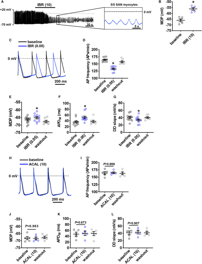
Figure 8. Effects of ibrutinib and acalabrutinib on spontaneous action potential (AP) morphology in isolated sinoatrial node (SAN) myocytes.

A, Representative spontaneous AP recordings demonstrating that 10 µmol/L of ibrutinib (IBR (10)) fully supresses AP firing in SAN myocytes. Recording is representative of the response in 5 SAN myocytes. B, Summary of the effect of IBR (10) on maximum diastolic potential (MDP) in SAN myocytes. *P<0.05 vs baseline by Student t test; n=5 SAN myocytes from 3 mice. C, Representative spontaneous SAN APs at baseline and after application of 0.05 µmol/L of ibrutinib (IBR (0.05)). D through G, Summary of the effects of IBR (0.05) on SAN AP frequency (D), MDP (E), APD at 50% repolarization (APD50) (F), and diastolic depolarization (DD) slope (G). For panels D–G: *P<0.0001 vs baseline by mixed effects analysis with a Tukey post hoc test; n=11 SAN myocytes from 6 mice. H, Representative spontaneous SAN APs at baseline and after application of 10 µmol/L of acalabrutinib (ACAL (10)). I through L, Summary of the effects of ACAL (10) on SAN AP frequency (I), MDP (J), APD50 (K), and DD slope (L). For panels I–L: data were analyzed by 2‐way repeated measures ANOVA with a Tukey post hoc test; n=5 SAN myocytes from 5 mice.
