Fig. 5.
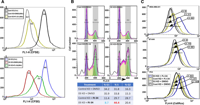
DHHC3 ablation enhances PARP inhibitor (PJ-34) effects. a The extent of CFSE staining decrease is used to assess proliferation of MDA-MB-231 cells that were DHHC3-ablated (D3 KD; bottom panel) or not ablated (Cont KD; top panel) using specific siRNA. Cells were CFSE loaded (for 36 h) and then either not further incubated (0 h; black, blue curves) or treated with DMSO (gray, red curves) or 10 µM PJ-34 (yellow, green curves) for an additional 48 h. b Propidium iodide fluorescence is used for cell cycle analysis of MDA-MB-231 cells transfected with control and DHHC3 siRNA, and treated with DMSO or PJ-34 (20 µM) for 24 h. c CellRox fluorescence is used to assess oxidative stress in cells (MDA-MB-231, top panel; BT-549, middle panel; PC3, bottom panel) that were DHHC3-ablated (D3 KD) or not ablated (Cont KD) using siRNA, and treated with either DMSO or PJ-34 for 24 h in complete media with 5% serum

