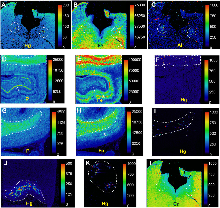
Fig 4. LA-ICP-MS in Parkinson’s disease (PD2).
(A) Particulate mercury is seen in both locus ceruleus nuclei (outlined) where numerous neurons were autometallography-positive. (B) Particulate iron deposits are seen in both locus ceruleus nuclei (outlined) in the posterior pons. Linear deposits of iron in the pontine white matter (eg, arrow) are probably from red blood cells in blood vessels. (C) Particulate aluminium is seen in both locus ceruleus nuclei (outlined). (D) A normal high nuclear density shown by the phosphorus image is present in the hippocampal white matter (top, outlined) as well as in the dentate gyrus (arrow). (E) A large amount of iron is present in the hippocampal white matter (outlined), and in grey matter adjacent to the dentate gyrus (arrow, dark blue line). (F) Particulate mercury is seen in the hippocampal white matter (eg, arrow) where oligodendrocytes were autometallography-positive. (G) The normal high nuclear density of the frontal white matter (outlined) is shown in the phosphorus image. (H) The frontal white matter contains more iron than the frontal cortex, where most iron is in the deeper cortical layers (*) adjacent to the white matter. (I) Particulate mercury is present in the frontal white matter where oligodendrocytes were autometallography-positive. (J) Speckled mercury is present in the lateral geniculate nucleus where neurons were autometallography-positive. (K) Mercury is seen in pontine facial motor neurons, which were autometallography-positive. (L) Chromium is widespread in the posterior pons, without particular accumulation in the locus ceruleus (outlined). Scale = counts per second (proportional to abundance).

