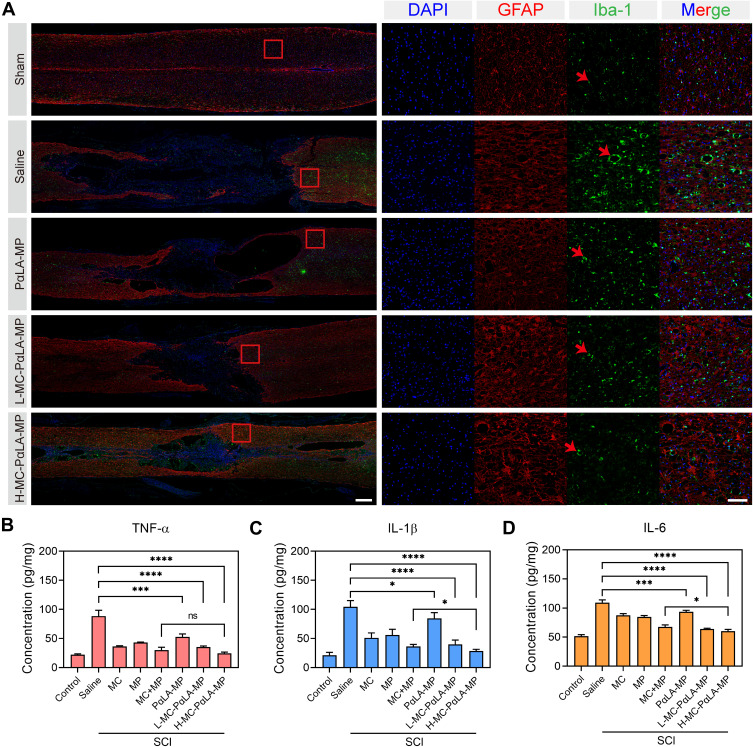
Figure 6.
Treatment of TSCI rats with intravenous MC-PαLA-MP NPs significantly reduced microglial activation, astrocyte activation, and production of pro-inflammatory cytokines at the injury site. (A) Immunofluorescence images of activated astrocytes (glial fibrillary acid protein-positive, red) and microglia (Iba1-positive, green) in the injured spinal cord. Low magnification images (2×) are on the left, and higher magnification images (10×) of the outlined areas are on the right. Scale bars are 500 and 100 μm, respectively. Arrows indicated the pathological characteristics. (B–D) Immunoexpression of (B) TNF-α, (C) IL-6, and (D) IL-1β in the spinal cord of TSCI rats receiving the indicated treatments. ****p < 0.0001; ***p < 0.001; *p < 0.05.
Abbreviation: ns, no significant difference.

