. 2021 Dec 24;10:e50324. doi: 10.7554/eLife.50324

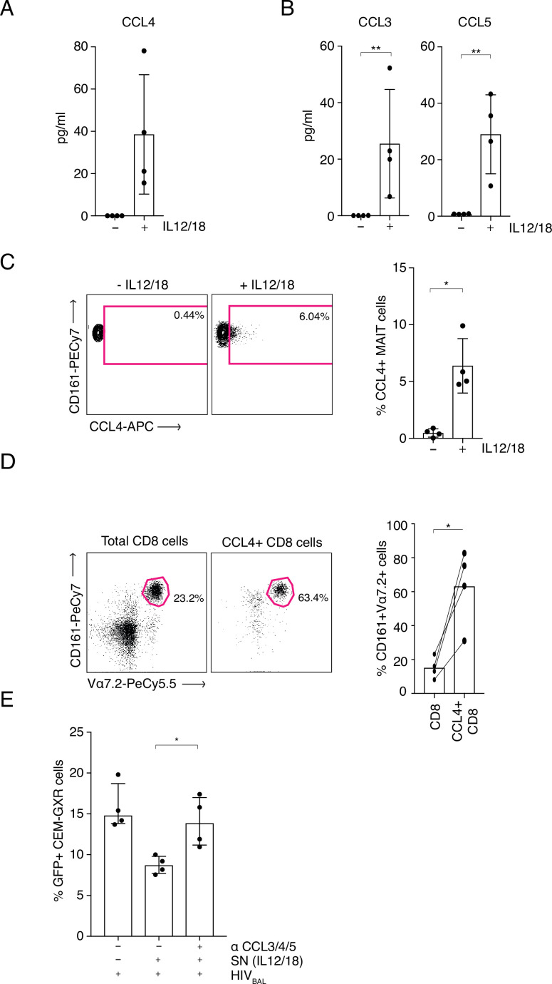
Figure 4. MAIT cell derived antiviral restriction factors are essential for suppressing HIV-1 in vitro.

(A) MAIT cells were FACS-sorted and the CCL4 (MIP-1β) concentration was measured in the supernatants by ELISA after 20 hr post stimulation with IL-12/18. (B) MAIT cells were FACS-sorted and the concentrations of CCL3 (MIP1α) and CCL5 (RANTES) were measured in the supernatants by cytometric bead array (CBA) after 20 hr post stimulation with IL-12/18. (C) Representative FACS-plots (left) and bar plots (right) depicting the expression of CCL4 by MAITs after incubation with IL-12/18 for 20 hr. MAIT cells were identified as CD161++ Vα7.2+ cells within MACS-enriched CD8s. (D) Representative FACS dot plots (left) and bar plots (right) showing the percentage of MAIT cells as identified by co-expression of Vα7.2 with high levels of CD161 within MACS-enriched CD8s and within all CCL4-expressing CD8 T cells from the same culture. CD8 T cells were stimulated with IL-12/18 for 20 hr. (E) Recovery of GFP-positive CEM-GXR cells following blocking of restriction factors (CCL3/4/5), after treatment with IL12/18 stimulated supernatant from CD8 cells and infection with HIVBAL. *p < 0.05, **p < 0.05, paired t-tests. Data were pooled from two independent experiments; error bars indicate the standard deviation.

Figure 4—source data 1. MAIT-cell-derived antiviral restriction factors are essential for suppressing HIV-1.
CCL4 (MIP-1β) concentration was measured in the supernatants by ELISA after 20 hr post stimulation with IL-12/18. (B) Concentrations of CCL3 (MIP1α) and CCL5 (RANTES) were measured in the supernatants by cytometric bead array (CBA) after 20 hr post stimulation with IL-12/18. (C) Expression of CCL4 by MAITs after incubation with IL-12/18 for 20 hr. MAIT cells were identified as CD161++ Vα7.2+ cells within MACS-enriched CD8s. (D) Percentage of MAIT cells as identified by co-expression of Vα7.2 with high levels of CD161 within MACS-enriched CD8s and within all CCL4-expressing CD8 T cells from the same culture. CD8 T cells were stimulated with IL-12/18 for 20 hr. (E) GFP-positive CEM-GXR cells following blocking of restriction factors (CCL3/4/5), after treatment with IL12/18 stimulated supernatant from CD8 cells and infection with HIVBAL.

Figure 4.

Figure 4—figure supplement 1. TCR-induced expression of CXCL12/SDF-1 by activated MAIT cells.
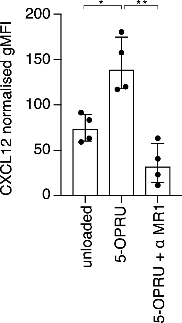
Figure 4—figure supplement 1.

(A) Intracellular expression (gMFI) of CXCL12 is increased with addition of 5-OPRU and decreased with a blocking antibody against MR1 (α MR1). Data points are biological replicates. Bar plot shown as mean and standard deviation. *p < 0.05, **p < 0.01; two-tailed t-tests.
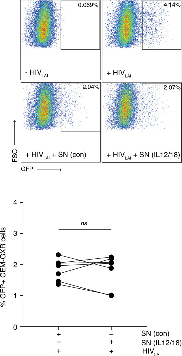
Figure 4—figure supplement 2. MAIT cells do not inhibit infection by a CXCR4 tropic virus.

Figure 4—figure supplement 2.

(A) Representative FACS plots showing infection of CEM-GXR cells with the CXCR4 tropic virus HIVLAI following pre-treatment with unstimulated or IL-12/18-stimulated MAIT cell supernatants. B. Bar graph showing HIVLAI infection of CEM-GXR cells after incubation with unstimulated or IL-12/18-stimulated MAIT cell supernatants. Data points are biological replicates. ns = not significant, paired two-tailed t-test.