FIG 1.

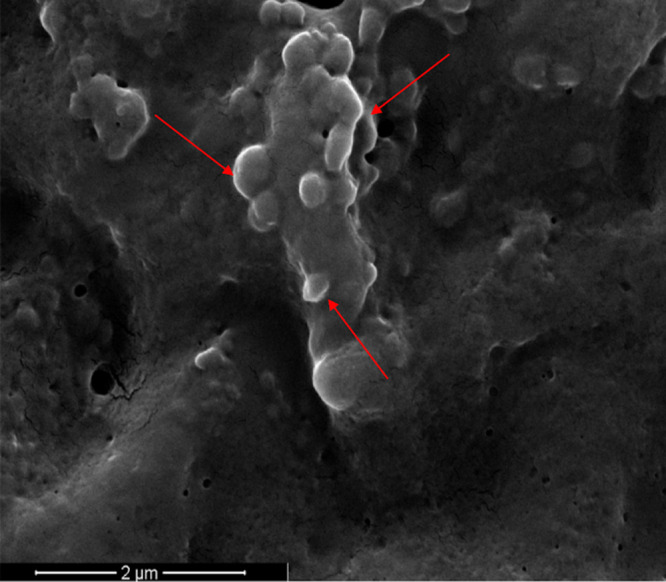
Scanning electron microscopy (SEM) image of S. coelicolor hyphae. Image was acquired after 6 days of bacterial growth in a liquid medium. Red arrows indicate some of the emerging MVs on the surface of bacterial hyphae.

Scanning electron microscopy (SEM) image of S. coelicolor hyphae. Image was acquired after 6 days of bacterial growth in a liquid medium. Red arrows indicate some of the emerging MVs on the surface of bacterial hyphae.