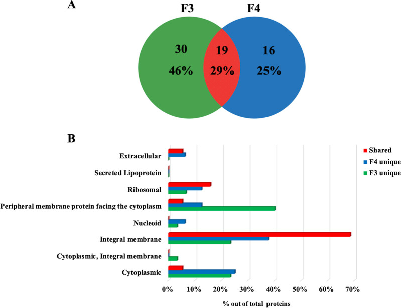
FIG 6.
Luminal proteins of S. coelicolor MVs. (A) Venn diagram showing the number of luminal proteins present in F3 and F4 MVs and the number that coincided. Percentage values refer to the total number of all identified proteins. (B) Predicted subcellular localization of proteins exclusively present in F3 or F4 MVs (“F3 unique” and “F4 unique”) or shared between the F3 and F4 MVs (“shared”). Percentage values refer to the total number of proteins identified in each fraction or shared.

