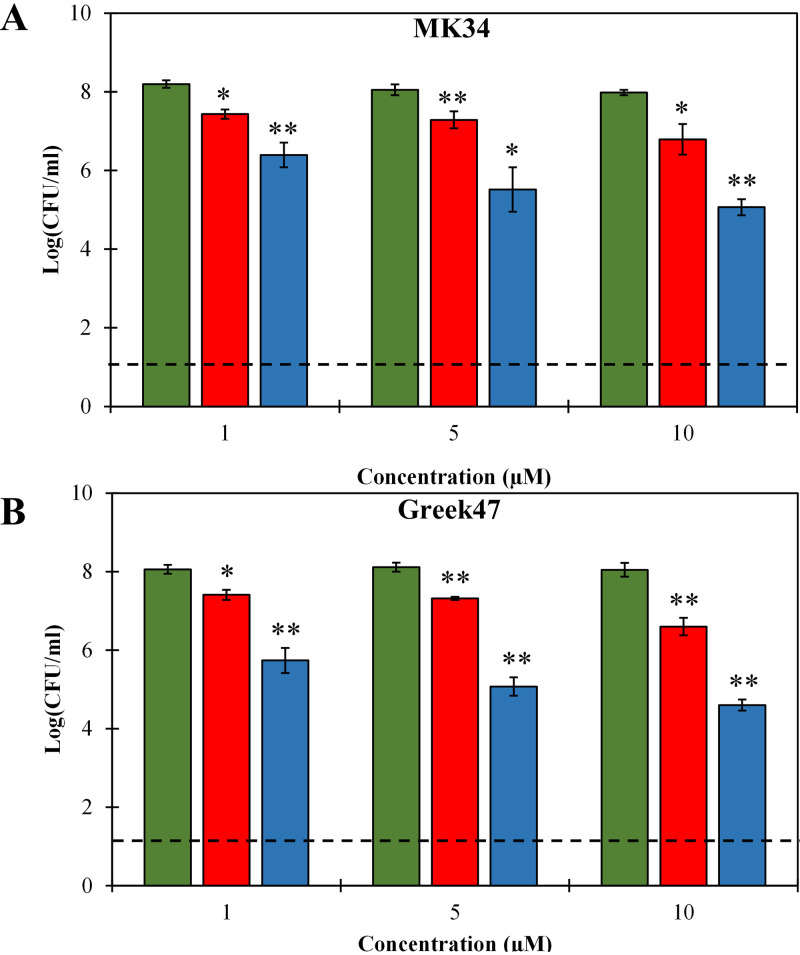
FIG 4.
Dose-dependent antibacterial activity of LysMK34 (red bars) and eLysMK34 (blue bars) against A. baumannii MK34 (A) and A. baumannii Greek47 (B) cells in the stationary growth phase with high intracellular osmotic pressure (suspended in 20 mM HEPES-NaOH, pH 7.4; ∼108 CFU/ml). The cells were exposed to either 1, 5, or 10 μM protein. Green bars show the untreated cells. The numbers of surviving bacteria are expressed as log numbers of CFU per milliliter after 120 min of exposure. Each value represents the mean ± standard deviation from three independent replicates. Asterisks represent statistical differences from values for buffer-treated cells (Student's t test; *, P < 0.05, **; P < 0.01; ***, P < 0.001). The dashed line represents the limit of detection (10 CFU/ml).

