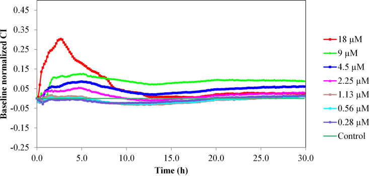
FIG 6.
Variation in the normalized cell index (CI) of HaCaT monolayers treated with different concentrations of eLysMK34 (0.28 to 18 μM). eLysMK34 was added to the RTCA wells after the HaCaT epithelial cells had formed a monolayer (reached after 20 h of incubation and indicated by a plateau in the cell index). The time point of eLysMK34 addition corresponds to time point zero. Subsequently, the cell index was monitored for 30 h upon addition of eLysMK34. Normalization of data was performed at 10 min after protein addition, with respect to the CI observed in the control sample (value 0 in the graph). Values represent means ± standard deviations from three replicates.

