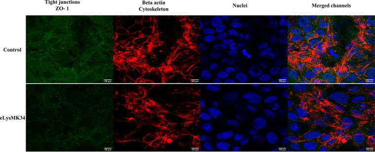
FIG 7.
Fluorescence images obtained by CLSM of HaCaT cells after 6 h of incubation with eLysMK34 (18 μM). As a control, cells were incubated without protein. The tight junctions, specifically, zonula occludes (ZO-1), were labeled with anti-ZO-1-Alexa Fluor 488 (green). The actin of the cytoskeleton was detected by labeling with Alexa Fluor 568 phalloidin probe (red), and the nuclei were labeled with DAPI (blue). The scale bars measure 10 μm.

