Figure 8.

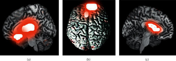
RS-fMRI images of a patient with abnormal mood. (a–c) The images in sagittal plane, transverse section, and coronal plane, respectively. The red fluorescence marked the abnormal high signal performances.

RS-fMRI images of a patient with abnormal mood. (a–c) The images in sagittal plane, transverse section, and coronal plane, respectively. The red fluorescence marked the abnormal high signal performances.