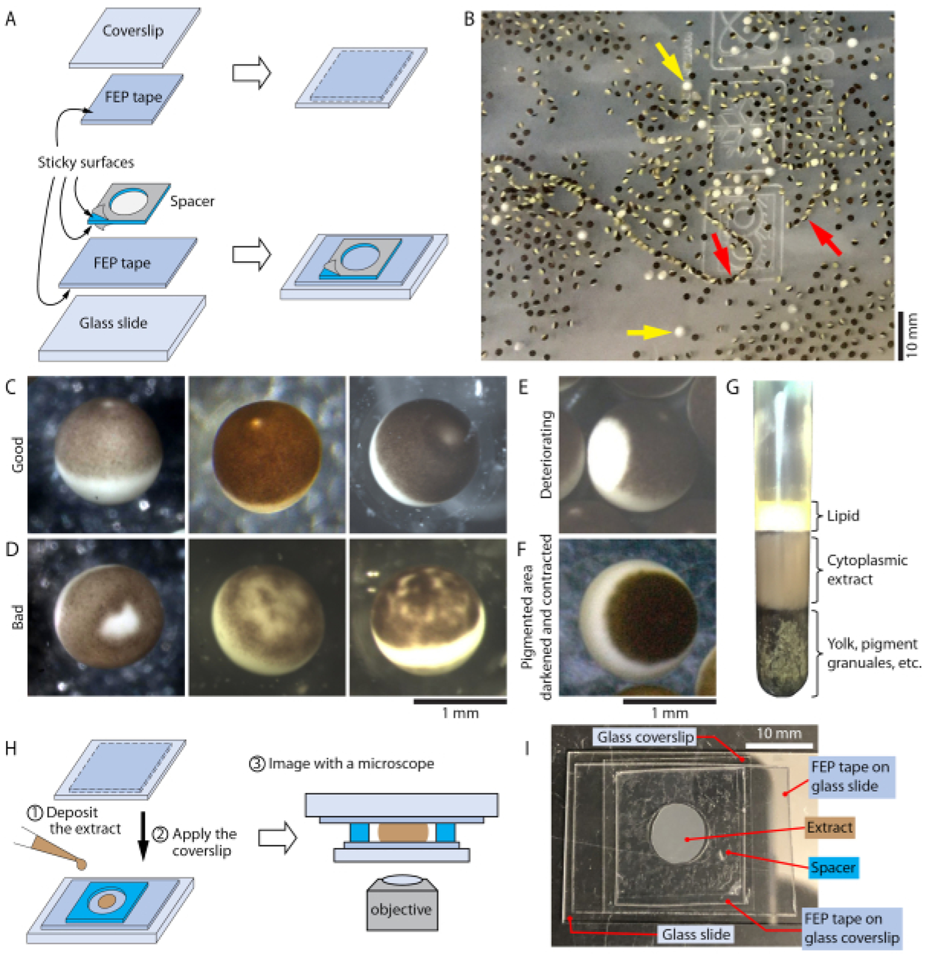
Figure 1: Schematics and photos related to the experimental procedure.

(A) Schematic diagram for preparing FEP tape-coated glass coverslips and slides. (B) Xenopus laevis eggs deposited in egg laying buffer, with examples of poor-quality eggs indicated by arrows. Yellow arrows, examples of eggs that look like white puffy balls. Red arrows, examples of eggs that appear in a string. (C) Examples of Xenopus laevis eggs with normal appearance. (D) Examples of poor-quality eggs with irregular or mottled pigment. (E) A deteriorating egg with a growing white region. (F) An egg that shows darkened and contracted pigmented area, possibly due to parthenogenetic activation. (G) The layers formed by ruptured Xenopus laevis eggs after the 12,000 × g centrifugation in step 2.11. (H) Schematics for preparing extract imaging chamber in step 2.15. (I) A photo of a prepared imaging chamber with an egg extract inside. (C) and (D) share the same scale bar at the bottom of (D). (E) and (F) share the same scale bar at the bottom of (F). The scale bars in (B) (C) (D) (E) (F) and (I) are approximate.
