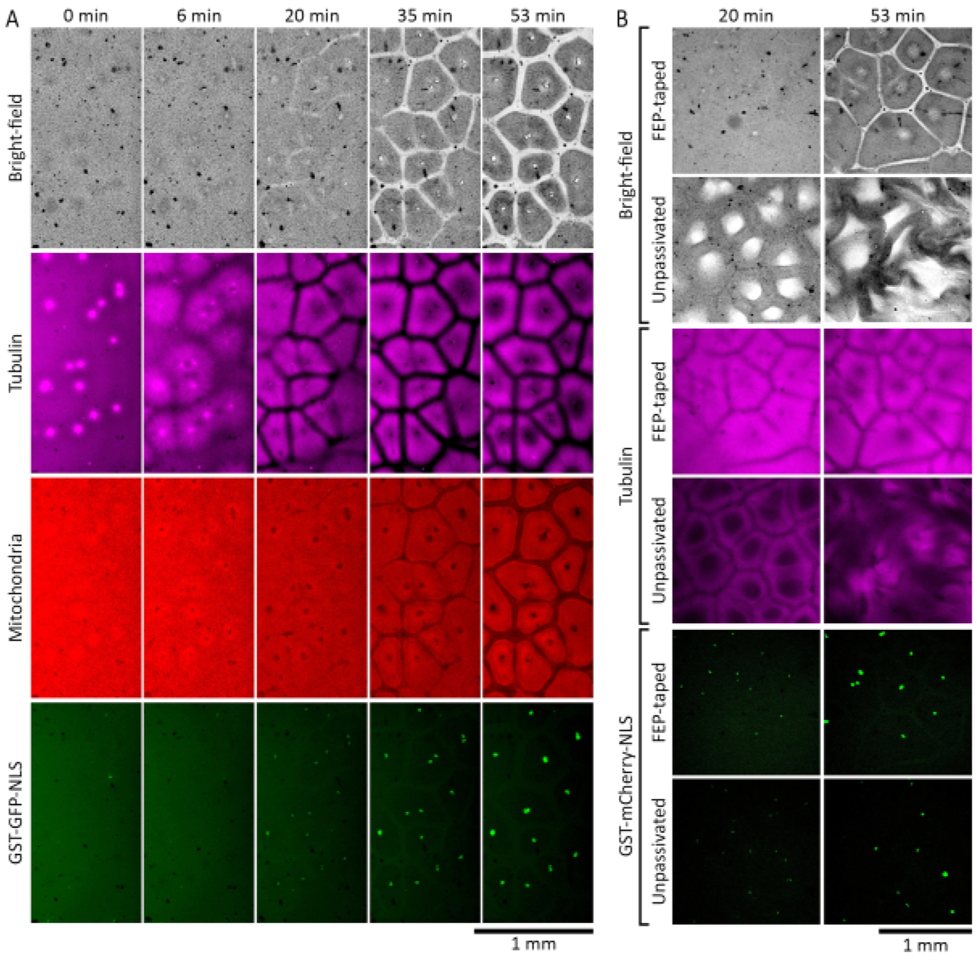
Figure 2: Interphase-arrested egg extracts self-organize into cell-like compartments.

(A)Time-lapse montage of self-organized pattern formation in a thin layer (120 μm) of interphase-arrested Xenopus laevis egg extract. The extract was supplemented with 27 nuclei/μL of demembranated Xenopus laevis sperm nuclei to allow reconstitution of the interphase nuclei. Microtubules were visualized by HiLyte 647-labeled tubulin (shown in magenta), mitochondria by MitoTrackerRed CMXRos (shown in red), and nuclei by GST-GFP-NLS (shown in green). (B) Self-organized pattern formation in interphase-arrested Xenopus laevis egg extracts placed in chambers with and without FEP-tape covered glass surfaces. The extracts were supplemented with 27 nuclei/μL of demembranated Xenopus laevis sperm nuclei to allow reconstitution of the interphase nuclei. Microtubules were visualized by HiLyte 488-labeled tubulin (shown in magenta) and nuclei by GST-mCherry-NLS (shown in green).
