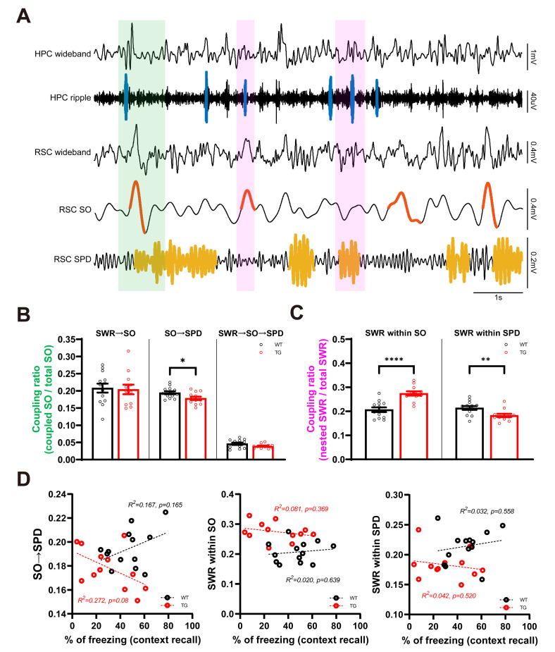
Fig. 5.
Altered sleep oscillation coupling between HPC and RSC in AD mice. (A) Example traces of HPC and RSC during SWS. Sharp wave ripple is marked on HPC ripple band (100~250 Hz) filtered trace with blue line. Cortical slow oscillation (SO) and spindle (SPD) events are labeled with red and yellow line respectively on filtered RSC channel (RSC SO : RSC 0.5~4 Hz filtered trace, RSC SPD : RSC 7~15 Hz filtered trace). Note the serial event of SWR-SO-SPD (green box) and co-occurrence of SWR within SO and SPD (magenta box). (B) Sequential appearance of SWR-SO and SWR-SO-SPD is unchanged, but SO-SPD pair event is reduced in TG mice (*p<0.05, Student’s t-test). (C) Co-occurrence ratio of SWR-SO and SWR-SPD is altered in TG mice (**p<0.01, **p<0.0001, Student’s t-test). (D) No correlation between changed HPC-RSC sleep oscillation coupling properties and freezing level in context recall.

