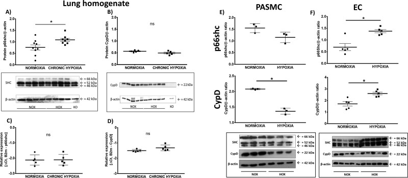
Figure 1.
Expression of p66shc and cyclophilin D (CypD) in lung tissue (A–D) after exposure to chronic hypoxia (10% O2; 4 weeks), and pulmonary arterial smooth muscle cells (PASMCs; E) or endothelial cells (ECs; F) of wild-type (WT) mice after a 5-day exposure to hypoxia. (A) Protein expression was determined with an antibody against SH2 containing proto-oncogene (shc) proteins p66shc, p52shc, and p46shc. P66shc expression was compared to β-actin expression. Lung homogenate from p66shc−/− mice (KO) was used as negative control (n = 9, from two western blots, one n excluded after outlier analysis). A representative western blot is shown. (B) Protein expression of CypD compared to β-actin expression. Lung homogenate from CypD−/− mice was used as negative control (n = 5). A representative western blot is shown. (C) Expression of p66shc mRNA compared to the expression of the reference gene (β2-microglobulin) in lung homogenate of WT mice (n = 5). (D) Expression of CypD mRNA compared to the expression of the reference gene (β2-microglobulin) in lung homogenate of WT mice (n = 5). (E) Protein expression in PASMCs determined with an antibody against cyclophilin D or SH2 containing proto-oncogene (shc) proteins p66shc, p52shc, and p46shc. Expression was compared to β-actin expression. n = 3 individual cell isolations. A representative western blot is shown. (F) Protein expression in ECs determined with an antibody against cyclophilin D or SH2 containing proto-oncogene (shc) proteins p66shc, p52shc, and p46shc. Expression was compared to β-actin expression. n = 5 individual cell isolations, one n excluded for quantitative analysis according to outlier analysis. A representative western blot is shown. * P < 0.05, determined by t-test. Data are shown as the mean ± SEM after outlier analysis.

