Figure 2.
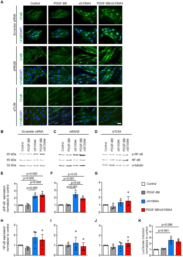
Oligomeric S100A4 activates SMCs through TLR4-dependent NF-κB pathway. Immunofluorescence staining (A) and representative Western blots for NF-κB and pNF-κB (B–D) followed by quantification (E–J) after transfection of S-SMCs with scramble siRNA, siRAGE, or siTLR4 followed by treatments with PDGF-BB, oS100A4, or PDGF-BB/oS100A4 for 1 h. Note that only by silencing of TLR4, oS100A4 and PDGF-BB/oS100A4-induced NF-κB activation is prevented (A, D, G, and J). In (B–D), α-tubulin is used as a housekeeping protein. In (A), nuclei are stained in blue by DAPI. Luciferase promoter reporter assay (K) after transfection of S-SMCs with pNFκB Tluc16-DD vector followed by treatments with PDGF-BB, oS100A4, or PDGF-BB/oS100A4 for 1 h. Note that promoter is activated after the treatment with oS100A4 and PDGF-BB/oS100A4. Bar = 25 µm, n = 3 biological replicates for each experiment. Comparisons were performed by using one-way ANOVA [E, F = 18.36, F(DFn, DFd) = 0.4488 (3,8); F, F = 16.63, F(DFn, DFd) = 1.201 (3,8); G, F = 1.021, F(DFn, DFd) = 0.6769 (3,8); H, F = 1.504, F(DFn, DFd) = 1.012 (3,8); I, F = 0.1510, F(DFn, DFd) = 0.7588 (3,8); J, F = 0.5399, F(DFn, DFd) = 0.5658 (3,8); K, F = 11.66, F(DFn, DFd) = 1.994 (3,8)].

