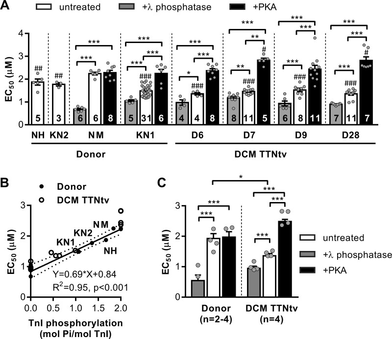
Figure 2.
Ca2+ sensitivity of force in cardiac myofibrils and its modulation via TnI phosphorylation. (A) The graph shows the concentrations of Ca2+ required for half-maximal force for donor and DCM patient hearts with truncating mutations in the TTN gene. Cardiac myofibrils were treated with PKA and λ phosphatase to fully phosphorylate and dephosphorylate TnI, respectively. Statistical analysis was performed using one-way ANOVA with Fisher’s least significant difference test. #P < 0.05, ##P < 0.01, and ###P < 0.001 vs. donor NM. *P < 0.05, **P < 0.01, and ***P < 0.001 vs. no treatment or other treatment. Numbers on bars indicate number of myofibril samples. (B) Correlation between myofilament Ca2+ sensitivity and TnI phosphorylation. The EC50 for Ca2+ required for half-maximal force responses in untreated, PKA-, and λ phosphatase-treated cardiac donor myofibrils (closed dots) are plotted against TnI phosphorylation level. The solid line is a least-squares linear regression line with 95% confidence interval (dotted line). Pearson correlation coefficient r = 0.91, n = 8 donor hearts (four untreated, two PKA-treated and two λ phosphatase-treated). The open circles are for DCM myofibrils (untreated, PKA-, and λ phosphatase-treated). (C) The EC50 values for the combined group of DCMs with TTNtv vs. healthy donors. Sarcomere length was 2.2 µm. Statistical analysis was performed using linear mixed model. Bars show estimated marginal means ± SE. Grey circles represent mean values of individual heart samples. *P < 0.05 and ***P < 0.001. Measurements were performed at 17°C.

