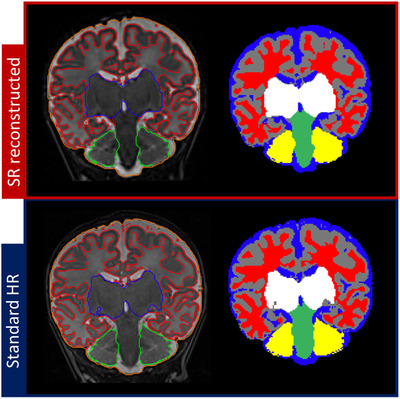
FIGURE 8.

Compared segmentations of standard 2‐dimensional high‐resolution image versus super‐resolution reconstructed 3‐dimensional volume of one subject (labels in red, white matter; gray, gray matter; blue, cerebrospinal fluid; white, basal ganglia; green, brain stem; yellow, cerebellum)
