
FIGURE 1.
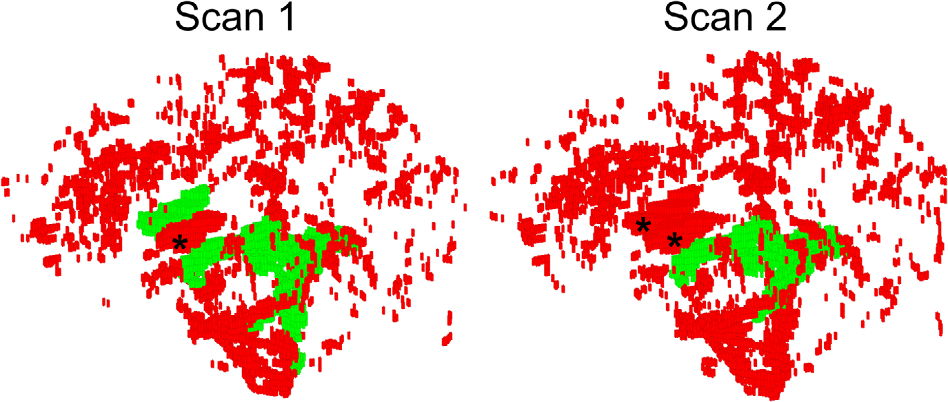
Volumetric rendering of the “global” cerebrospinal fluid (CSF) volume (red) and its overlap with “ventricular CSF” (green) reconstructed from a scan-rescan data of the same healthy volunteer. Note that elimination of the unconnected components in ventricular CSF method resulted in complete erroneous elimination of the anterior horns of lateral ventricles (marked with “*”) in both segmentations, and introduced inconsistent ventricular masking. Furthermore, large volumes of CSF spanning the brain volume are completely discarded during the morphological filtering. While ventricular CSF segmentation demonstrates noticeable variations between scans, the global CSF mask is stable. See Methods for further details