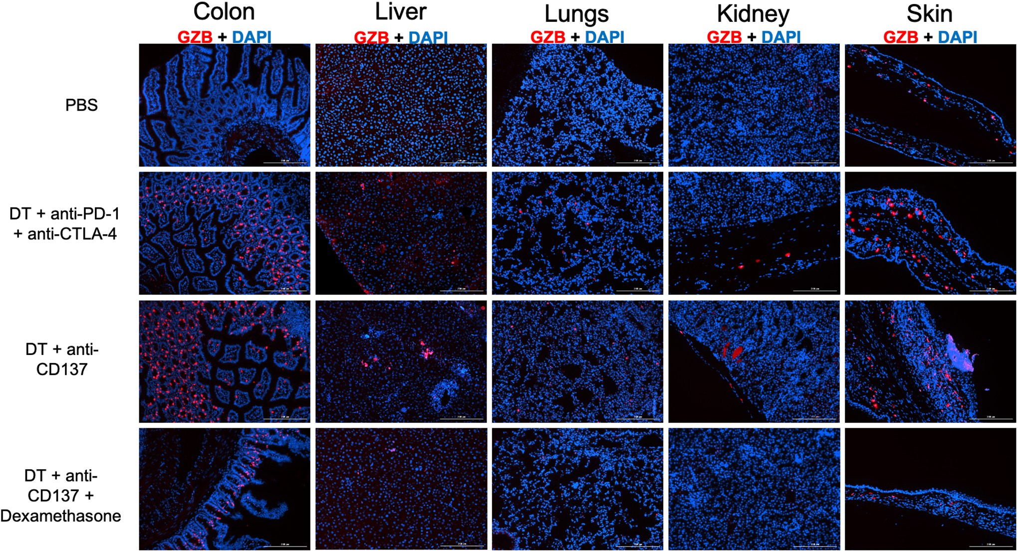
Figure 3. Immunofluorescence staining against granzyme B (in red) of major organs of different treatment groups reveal granzyme B presence in the groups injected with DT + immune checkpoint inhibitors.

Cell nuclei is stained with DAPI (blue). Magnification 10X.
