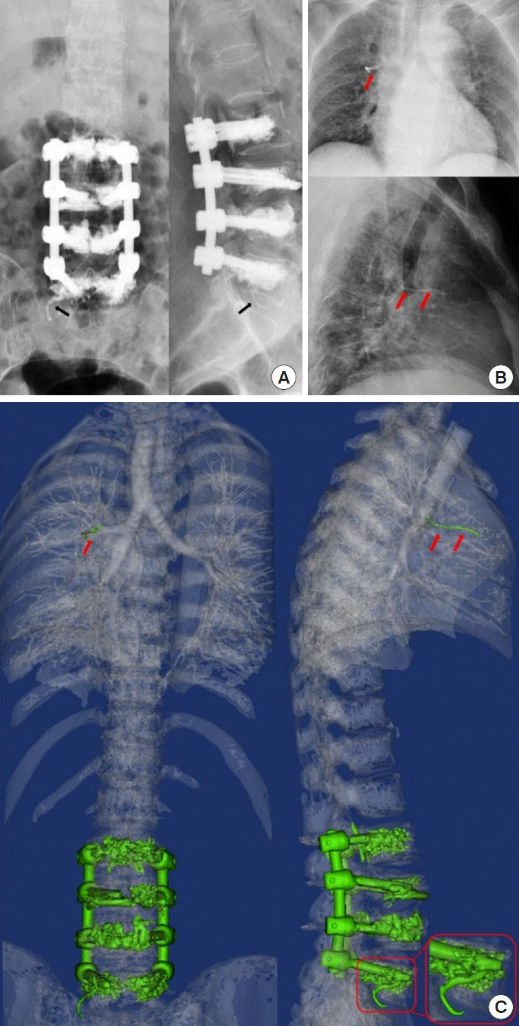
Fig. 2.

A 73-year-old female developed postoperative pulmonary cement embolism after cement-augmented pedicle screw instrumentation at the L2–5 level (case No. 28). (A) Anteroposterior and lateral digital radiographs showed curvilinear cement in the paravertebral venous plexus (black arrow). (B) Postoperative chest x-rays showed a linear-like, hyperdense cement embolism in the right lung (red arrow). (C) The zoomed region of the red box shows cement leakage into the paravertebral venous plexus at the L5 level. In addition, a cement embolism was found in the right pulmonary vascular tree (red arrows).
