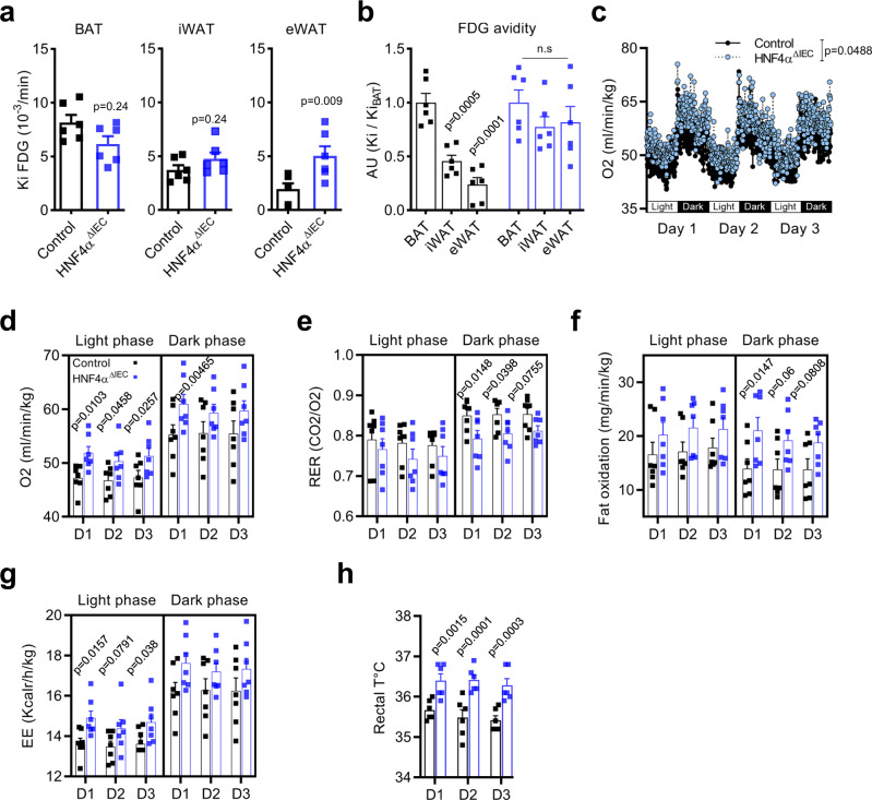
Fig. 4. Loss of intestinal epithelial HNF4A impacts WAT metabolism, fat oxidation, and energy expenditure in mice fed a HFD.
a PET estimates of metabolic rates based on FDG uptake (Ki) by BAT, iWAT, and eWAT from control (black squares) and HNF4AΔIEC mutant (blue squares) mice fed a HFD for 2 weeks (n = 6 biologically independent animals). Statistical comparisons were performed using two-tailed Mann–Whitney test. b Relative WAT FDG were reported to BAT FDG fractional uptake (n = 6) Statistical comparisons were performed using two-way ANOVA followed by uncorrected Fisher’s LSD test. c Oxygen consumption from indirect calorimetry of control (black circles) and HNF4AΔIEC mice (blue circles) fed a HFD for 2 weeks (n = 7) Statistical comparisons were performed using two-way ANOVA test. Averages of oxygen consumption (d), respiratory exchange ratio RER (e) fat oxidation (f), and energy expenditure (EE) (g) per daily phases for the last 3 days of HFD for control (black squares) and HNF4AΔIEC mice (blue squares) (n = 7) Statistical comparisons were all performed using two-way ANOVA followed by uncorrected Fisher’s LSD test. h Rectal temperatures of control (black squares) and HNF4AΔIEC mutant (blue squares) mice during the last 3 days of HFD (n = 6). Statistical comparisons were performed using two-way ANOVA followed by uncorrected Fisher’s LSD test. Data are presented as mean values ± SEM. Source data are provided as a Source Data file.

