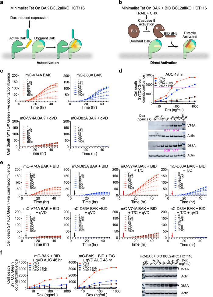
Fig. 3. BAK autoactivation and direct activation cooperate in cells.
a, b Schematic of minimalist BAK ± BID apoptotic pathway reconstituted in BCL2allKO HCT116 cells. c Apoptosis of BCL2allKO HCT116 cells reconstituted with Dox-inducible V74A and D83A mCherry-BAK (mC-BAK) monitored for 48 h by IncuCyte imaging uptake of cell-impermeable dye SYTOX Green. Data are presented as mean + SD of one representative from n = 3 experiments each of n = 3 technical replicates. Dox concentration (ng/mL) is inset. d AUC of kinetic traces in (c) and representative immunoblots from n = 2 independent experiments. The purple line indicates similar cell death induced at ~10-fold higher expression level for D83A compared to V74A. The ratio of background-corrected mC-BAK/actin is shown in purple for 50 ng/mL and 500 ng/mL Dox. e Apoptosis of BCL2allKO HCT116 cells reconstituted with Dox-inducible V74A and D83A mC-BAK monitored for 48 h by IncuCyte imaging uptake of cell-impermeable dye SYTOX Green. Culture medium ± qVD (black arrows) or TRAIL + CHX (T/C) ± qVD (red arrows) were added at 6 h after addition of Dox. Data are presented as mean + SD of one representative of n = 4 experiments each of n = 3 technical replicates. Dox concentration (ng/mL) is inset. f AUC of kinetic traces in (e) and representative immunoblots from n = 2 independent experiments after 24 h of incubation with Dox+qVD. BAK, arrowhead; *, nonspecific band.

