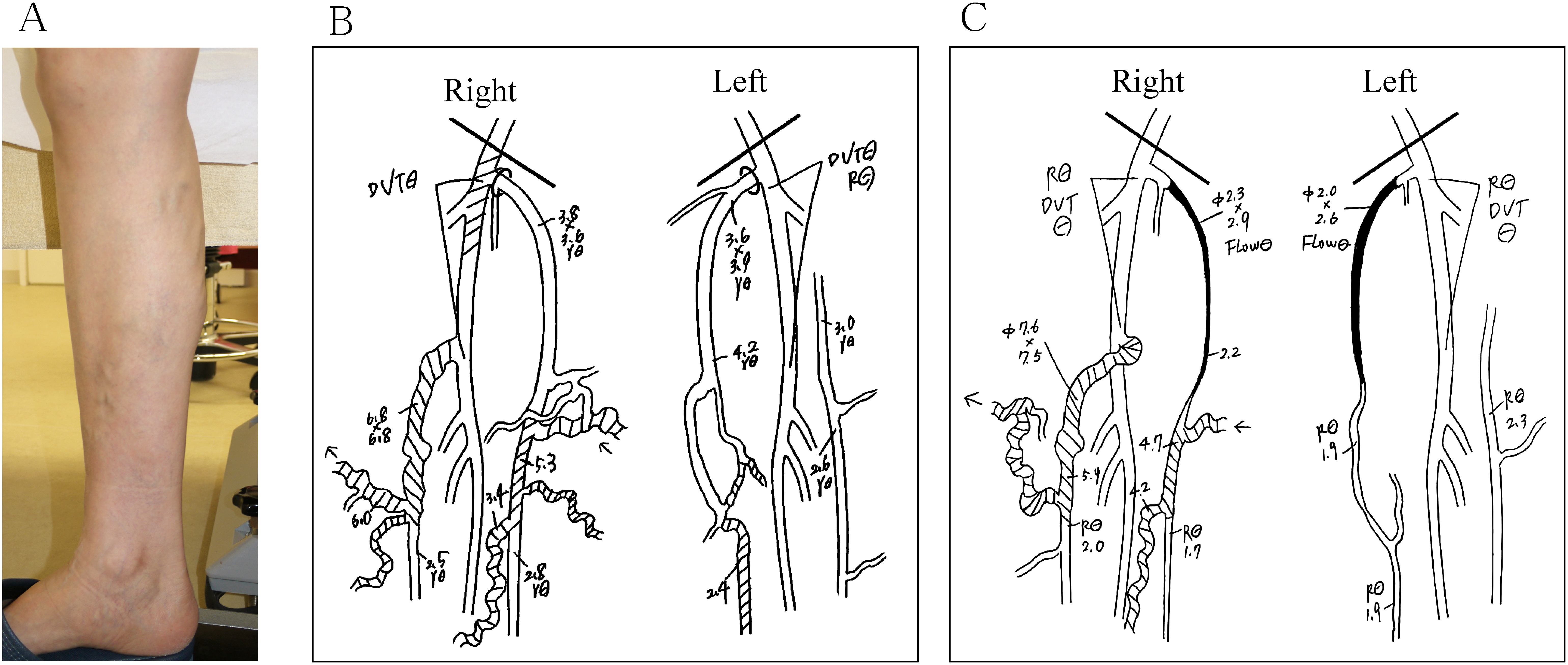
Fig. 2 A case of improper treatment.
(A) Macroscopic findings: Lower extremity varicose veins are seen on the medial side of the right lower limb. (B) Venous ultrasonography findings at the first examination: Valvular insufficiency is seen in the right SSV and femoral vein and left distal GSV. Dilatation is seen in the right SSV with a diameter of 6.8×6.8 mm. The shaded area indicates the range of valvular insufficiency. (C) Venous ultrasonography findings at the reexamination: Bilateral GSV is occluded by ETA. Valvular insufficiency and dilatation of the right SSV expanded to a diameter of 7.6×7.5 mm. SSV: small saphenous vein; GSV: great saphenous vein; DVT: deep vein thrombosis