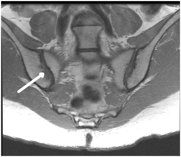
Fig. 1.

Subchondral resorption. Coronal oblique proton density intermediate-weighted image shows right sacroiliac joint subchondral resorption (arrow) demonstrating loss of the hypointense (dark) subchondral plate without defined erosion. Note that the left sacroiliac has a maintained subchondral plate (dark line).
