Fig. 1.
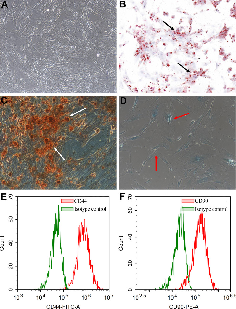
Primary culture and multiple differentiation of ADSCs. These spindle-shaped cells isolated from human adipose tissue adhered to the Petri dishes and formed a monolayer (A). ADSCs differentiated into adipocytes as lipids were accumulated in ADSCs culture. Black arrows indicate the presence of lipids droplets (B). ADSCs differentiated into osteoblasts as extracellular calcium phosphate deposits were observed in ADSCs culture. White arrows indicate the presence of osteogenic mineral deposition (C). ADSCs differentiated into chondroblasts as nodules were formed in ADSCs culture. Red arrows indicate the presence of nodules and inter chondrogenic nodule connections (D). ADSCs were positive for CD44 (E). ADSCs were positive for CD90 (F)

