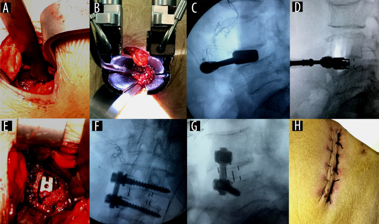
Figure 1. Surgical technique.
(A) A 3.0-cm centered skin incision was made in projection of the target segment; (B) the tubular retractor system was placed in the targeting disc; (C) the mold of the cage was tested and examined under a fluoroscope; (D) the cage was placed and examined under a fluoroscope; (E) the cage was directly observed in the intervertebral space; (F) intraoperative fluoroscopy in the anteroposterior position; (G) intraoperative fluoroscopy in the lateral position; and (H) the surgical wound after suturing.

