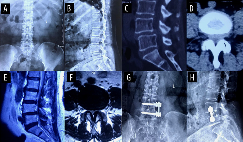
Figure 5. A typical case of oblique lateral interbody fusion with anterolateral screw fixation (OLIF+AF).
A 65-year-old woman had low back pain for 4 years, aggravated for 3 months. OLIF+AF was employed to treat lumbar spondylolisthesis at L4/5. (A) Preoperative anteroposterior radiograph. (B) Preoperative lateral radiograph shows L4 vertebral body I° anterior spondylolisthesis and loss of intervertebral disc height at L4/5. Before surgery, the anterior disc height (ADH) was 5.3 mm, the posterior disc height (PDH) was 4.9 mm, and the foraminal height (FH) was 13.2 mm. (C) Preoperative sagittal computed tomography (CT). (D) Preoperative cross-section CT shows disc herniation, obvious proliferation of the ligamentum flavum, and spinal canal stenosis at L4/5. (E) Preoperative sagittal magnetic resonance imaging (MRI). (F) Preoperative cross-section MRI shows disc herniation, obvious proliferation of the ligamentum flavum, and spinal canal stenosis at L4/5. (G) Postoperative anteroposterior radiograph. (H) Postoperative lateral radiograph shows the intervertebral cage at L4/5 was well in place, and the ADH, PDH, and FH were significantly increased. After surgery, the ADH was 10.4 mm, the PDH was 9.7 mm, and the FH was 18.3 mm.

