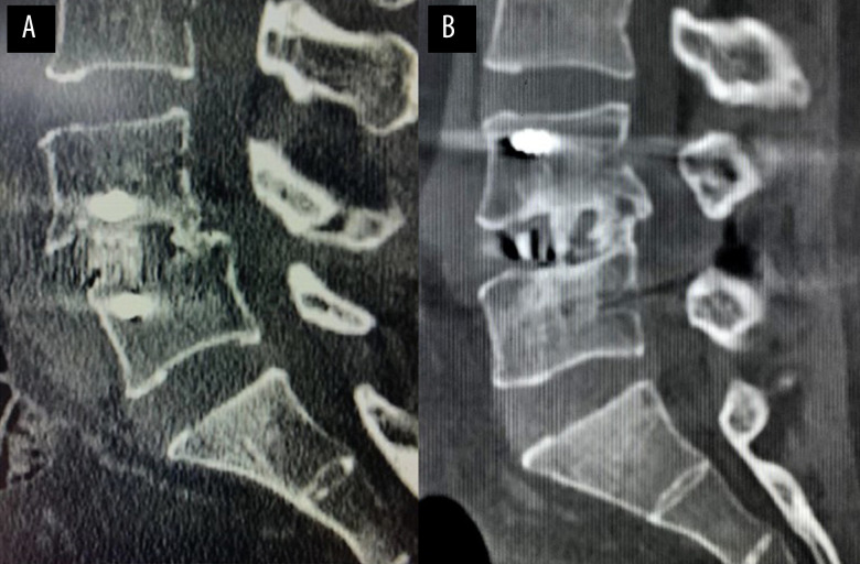
Figure 7. The typical case of postoperative fusion in the OLIF+AF and OLIF+PF groups.
(A) Postoperative sagittal computed tomography (CT) in OLIF+AF case shows intervertebral fusion with trabeculae reconstruction and no lucencies at the top or bottom of the graft at L4/5. (B) Postoperative sagittal CT in OLIF+PF case shows intervertebral fusion with trabeculae reconstruction and no lucencies at the top or bottom of the graft at L4/5.

