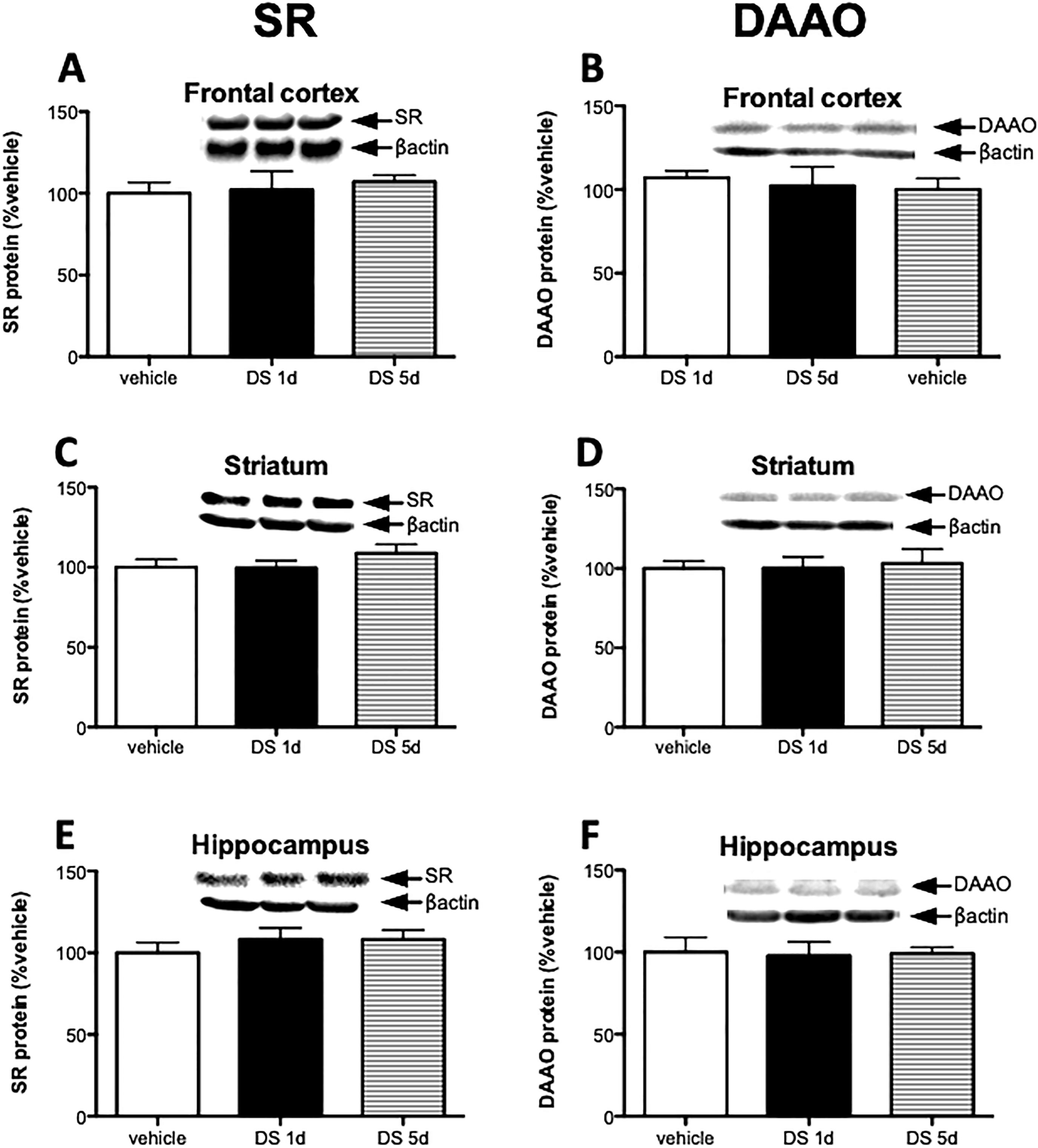
Fig. 4.

D-serine treatment does not regulate SR and DAAO expression. Mice were divided to 3 groups: vehicle (n = 6), 1 day of D-serine treatment (DS 1d; n = 5), and 5 days D-serine treatment (DS 5d; n= 5). Protein levels of SR (A, C, E) and DAAO (B, D, F) were measured in the frontal cortex (A, B), striatum (C, D) and hippocampus (E, F) of vehicle group (white bars), 1 day D-serine group (black bars) and 5 day D-serine group mice (stripe bars). Values are expressed as the optical density (OD) normalized to vehicle treated values (% vehicle). One-way ANOVA was used for analysis. All values represent the mean ± SEM.
