
Fig. 1.
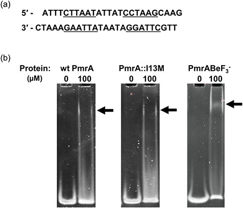
Defining the A. baumannii PmrA binding motif. (a) KpPmrA_25mer DNA sequence derived from the K. pneumoniae PmrA box (42). Inverted repeat sequences are underlined. (b) EMSAs showing binding of wt PmrA, PmrA::I13M and wt PmrA-BeF3− to 1 μM KpPmrA_25mer. Arrows indicate positions of mobility shifts.