
Fig. 5.
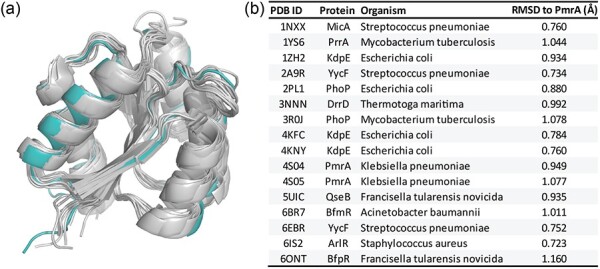
Structural alignment of A. baumannii PmrAN with other response regulators. (a) Structural alignment of our PmrAN structure to 16 other response regulator protein structures from the PDB. Acinetobacter baumannii PmrA is shown in teal and other response regulators are shown in grey. (b) Table showing RMSD values for alignment of each structure with A. baumannii PmrAN. A lower RMSD value indicates higher similarity between structures. Alignments were performed to chain A of each PDB structure. Values were calculated using PyMol.