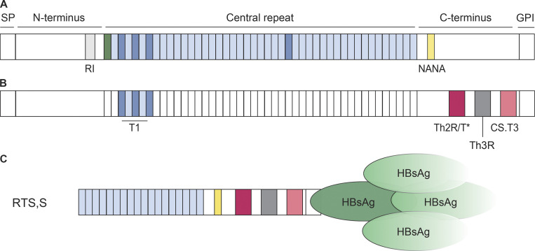
Figure 2.
Schematic representation of PfCSP and RTS,S. (A) PfCSP consists of a signal peptide (SP), an N-terminal domain with the RI, a central repeat region, and a C-terminal domain. It is anchored in the sporozoite membrane through a GPI anchor. The N-terminal domain is linked to the central repeat region, composed of repeating NANP motifs (light blue), via a junctional region with a single NPDP (green) followed by a small number of NANP and alternating NVDP (dark blue) motifs. Protective antibodies recognize these four aa motifs and a single NANA (yellow) motif in the C-terminal domain (Julien and Wardemann, 2019). (B) Known T cell epitopes in the junctional region (T1; Nardin et al., 1989) and the C-terminal domain (Th2R/T*, Th3R, CS.T3; Good et al., 1988; Guttinger et al., 1988; Sinigaglia et al., 1988a; Moreno et al., 1991) are highlighted in colors. (C) RTS,S consists of 18 NANP repeats and the complete PfCSP 3D7 C-terminal domain fused to HBsAg and complexed with free HBsAg in a 1:3 ratio.

