FIG 9.
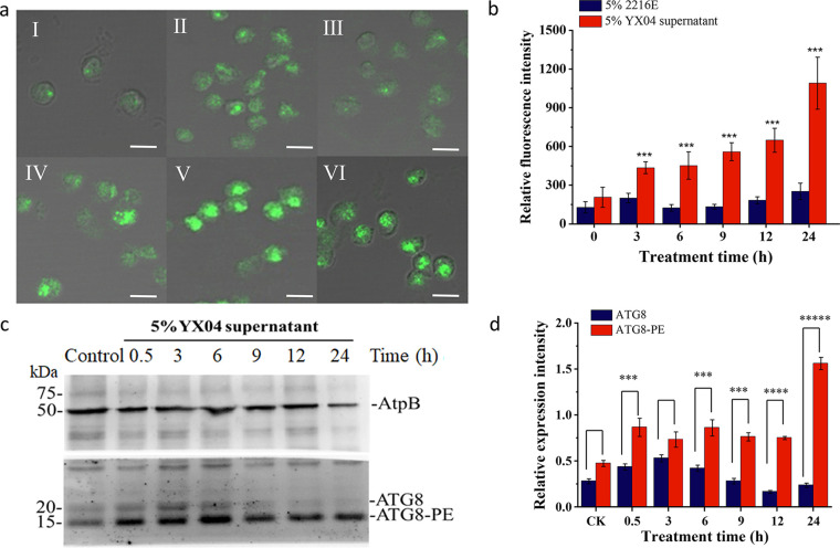
Detection of the autophagic flux in P. globosa cells treated with 5% YX04 supernatant. (a) Immunolocalization of ATG8 in P. globosa cells treated with 5% YX04 supernatant at 3 (II), 6 (III), 9 (IV), 12 (V), and 24 h (VI) and with the control 5% 2216E broth for 24 h (I), scale bar = 5 μm. (b) Relative immunofluorescence intensity of panel a. Thirty cells were selected in each sample to calculate the mean value; the error bars indicate the standard errors of 30 cells. (c) Western blotting (WB) of ATG8 protein expression in algal cells treated with 5% YX04 supernatant for different amounts of time. Anti-AtpB antibodies were used as the loading control. (d) The relative intensities of the WB bands were analyzed using ImageJ software. The y coordinate indicates the ratio of the gray value of the ATG8 band to the gray value of the band corresponding to the internal reference protein AtpB. Different sample collection time points are labeled on the x coordinate. *, P < 0.05; **, P < 0.01; ***, P < 0.001 compared with the control.

