FIG 6.
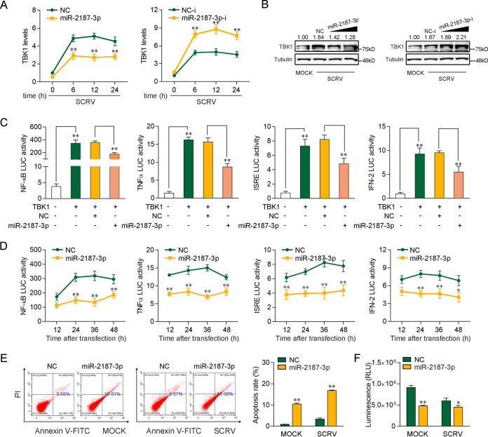
miR-2187-3p can negatively regulate TBK1-mediated antiviral signaling. (A) miR-2187-3p inhibits the mRNA level of endogenous TBK1 upon SCRV infection. qPCR assays were performed to determine the expression levels of TBK1 in MICs transfected with miR-2187-3p, NC, miR-2187-3p-i, or NC-i and infected with SCRV for different times. (B) miR-2187-3p suppresses the protein expression of endogenous TBK1 upon SCRV infection. MICs were cotransfected with miR-2187-3p or NC at different concentrations and miR-2187-3p-i or NC-i at different concentrations for 24 h. Then, the cells were treated with SCRV for 24 h, and the expression of TBK1 was determined by Western blotting. (C) miR-2187-3p inhibits TBK1-activated luciferase activity. EPC cells were cotransfected with NC or miR-2187-3p, TBK1 expression plasmid, and pRL-TK vector, along with the NF-κB, TNF-α, ISRE, or IFN-2 reporter gene, to investigate the regulatory effect of miR-2187-3p on NF-κB and IRF3 signals. (D) Relative luciferase activity of indicated reporters in EPC cells after cotransfection with NC or miR-2187-3p was determined. EPC cells were cotransfected with NC or miR-2187-3p, the TBK1 expression plasmid, and the pRL-TK vector, together with the NF-κB, TNF-α, ISRE, or IFN-2 luciferase reporter gene, and then the luciferase activity was measured over time, as indicated. (E) The effect of miR-2187-3p on cell apoptosis was analyzed by flow-cytometric cell apoptosis assays. The SCRV treatment was performed after transfection with NC or miR-2187-3p at 24 h. (F) Effect of miR-2187-3p on cell viability. MICs were transfected with NC or miR-2187-3p for 24 h and then infected with SCRV for 24 h. A cell viability assay was performed. Error bars indicate the SE for data from three independent experiments. **, P < 0.01, and *, P < 0.05, compared to results with the control.

