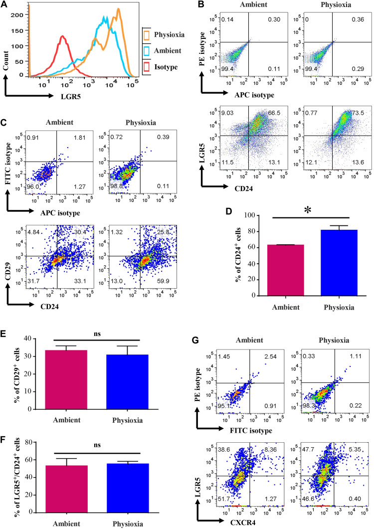
Fig. 6. Cells derived from mouse colon collected and processed under physioxia display distinct marker profile compared to that of cells in colon processed under ambient air.
Mouse colon was collected under physioxia, washed, and minced. A fraction was transferred to ambient air for processing, whereas the other fraction was processed under physioxia (n = 3, one-way ANOVA). (A) Intensity of LGR5 staining. (B) LGR5 and CD24 staining patterns of colonic cells under physioxia and ambient air. Only lineage-negative cells were gated for the analysis. (C) CD24/CD29 staining patterns of colonic cells. FITC, fluorescein isothiocyanate. (D) Percentage of CD24+ cells under physioxia and ambient air. CD24+ cells were higher under physioxia. (E) CD29 positivity did not differ between physioxia and ambient air. (F) Number of LGR5+ cells did not differ between physioxia and ambient air, although staining intensity was higher under physioxia. (G) Physioxia did not have an influence on CXCR4 positivity. *P < 0.05 by ANOVA.

