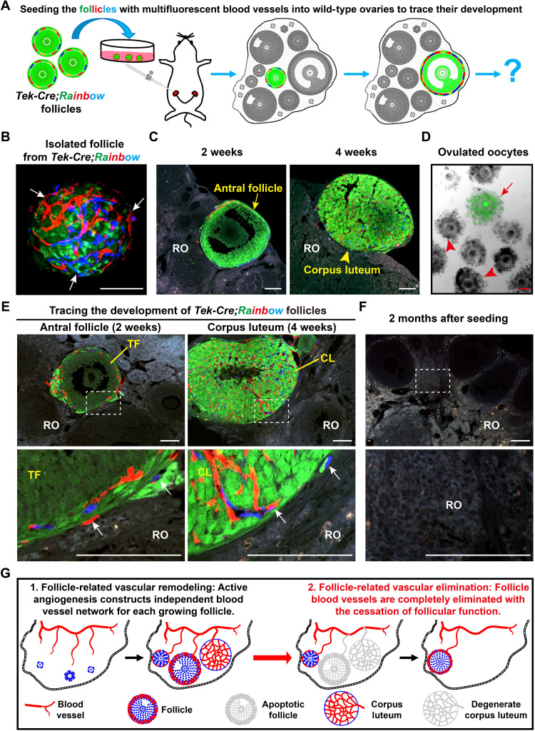
Fig. 3. Angiogenesis established an independent and temporary follicle vascular network in the adult ovaries.
(A) Illustration of the strategy used to trace the development of follicle blood vessel endothelial cells in vivo. GFs with fluorescent-labeled blood vessels were isolated from Tek-Cre;Rainbow females and were surgically seeded into the ovaries of wild-type recipient females for long-term tracing. (B) Before seeding, a single follicle from Tek-Cre;Rainbow ovaries showed red or blue fluorescent blood vessels (arrows) surrounding the GFP follicle. (C and D) The transplanted Tek-Cre;Rainbow follicles successfully developed to the antral stage and formed a CL in the recipient ovaries (ROs) and also ovulated the GFP oocytes from recipient females, showing the normal development of the seeded follicles. (E) Tracing the developmental fate of follicle blood vessels after transplantation. All fluorescent blood vessels (arrows) were limited to the surface of transplanted follicles and the CL, which were derived from transplanted follicles. (F) At 2 months after seeding, all fluorescent blood vessels were eliminated because of the exhaustion of transplanted follicles in the ROs (0 of 97). The transplanted window is indicated by the white frame. (G) Schematic of the fate of new follicular blood vessels formed via angiogenesis, showing the formation of an independent blood vessel network for each GF in the ovaries, and a complete elimination of follicle blood vessels with the cessation of follicle function. RO, recipient ovary; TF, transplanted follicle; CL, corpus luteum. Scale bars, 100 μm (B to E).

