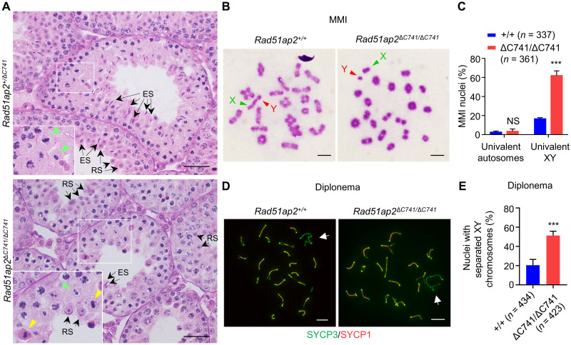
Fig. 2. Precocious separation of XY chromosomes in Rad51ap2 mutant spermatocytes.
(A) Testicular histology from 6-week-old WT and Rad51ap2 mutant mice. Green arrowheads indicate normal metaphase cells, yellow arrowheads indicate metaphase cells with unaligned chromosomes, and arrows indicate representative round spermatids (RSs) or elongating/elongated spermatids (ESs). Scale bars, 50 μm. (B) MMI spermatocytes stained with Giemsa. Chromosomes X and Y are indicated. Scale bars, 10 μm. (C) Frequencies of nuclei with XY chromosome or autosome dissociation in MMI spermatocytes. n, the number of MMI cells scored from four mice per genotype. NS, not significant. ***P < 0.001, two-way analysis of variance. (D) Representative spread spermatocytes in diplonema, stained for SYCP3 (green) and SYCP1 (red). Arrows indicate the X and Y chromosomes. Scale bars, 10 μm. (E) Frequencies of nuclei with XY separation in diplotene spermatocytes. n, the number of cells scored from at least two mice per genotype. ***P < 0.001, two-tailed Student’s t test.

