
Fig. 1. CM-RevDKO mice die from dilated cardiomyopathy.
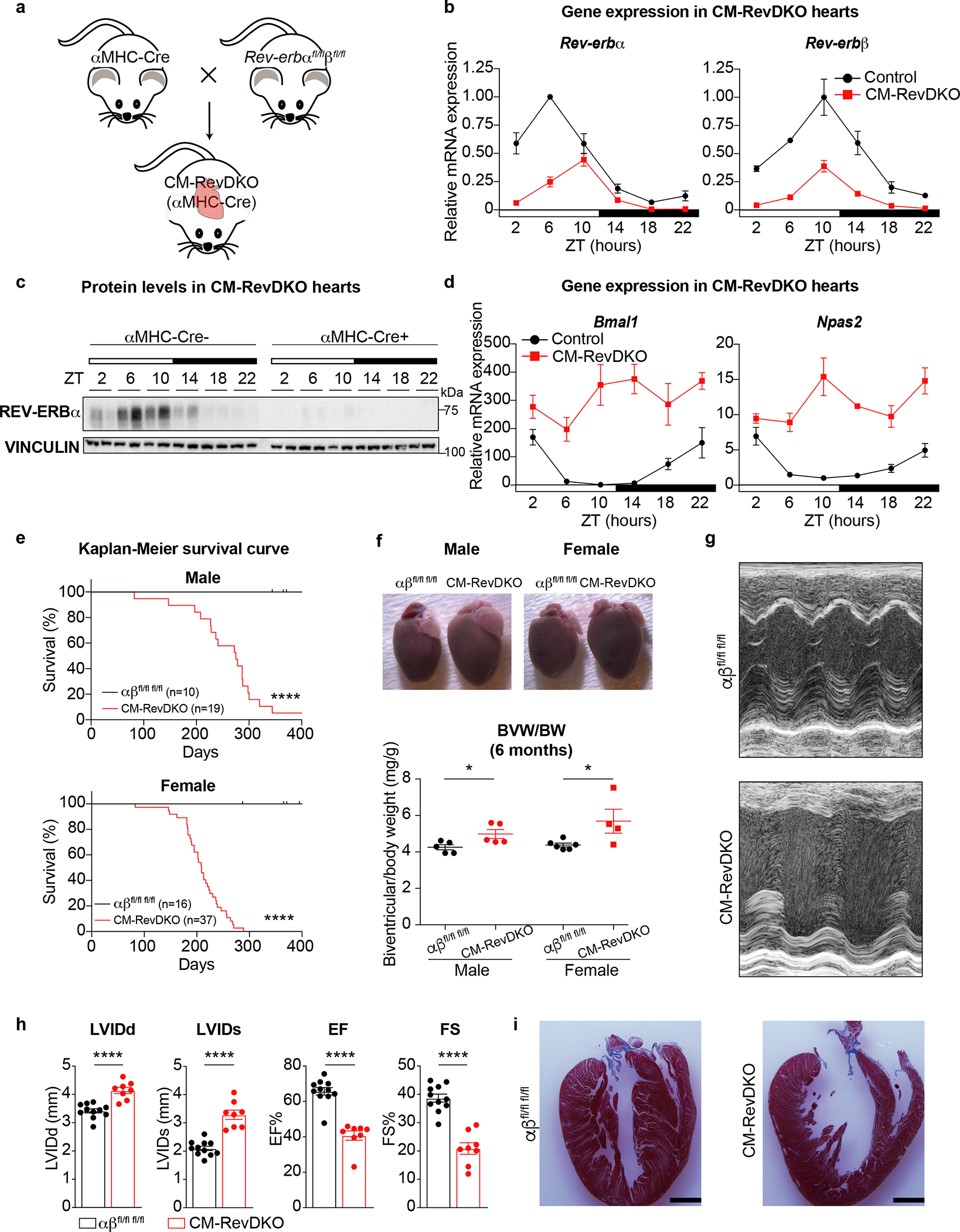
a, Cartoon depicting strategy for cardiomyocyte-specific Rev-erbα/β double knock out model (CM-RevDKO) generation by crossing cardiomyocyte-specific Cre-deleter line (αMHC-Cre)21 with a recently developed Rev-erbαff/βff mouse model20. b, Relative mRNA expression in hearts from 2-month-old male αMHC-Cre+ Rev-erbα/β double floxed (CM-RevDKO) vs littermate controls (αMHC-Cre-) (n = 3 hearts/genotype/timepoint except for n = 2 for ZT6 and ZT22 in control, ZT2 and ZT18 in CM-RevDKO, and n = 4 for ZT22 in CM-RevDKO). c, Immunoblot for REV-ERBα from control (αMHC-Cre-) vs CM-RevDKO (αMHC-Cre+) hearts from 2-month-old male mice (n = 2/timepoint/genotype). d, Relative REV-ERBα/β target gene mRNA expression in control vs CM-RevDKO hearts from 2-month-old male mice (n = 3 hearts/genotype/timepoint except for n = 2 for ZT6 and ZT22 in control, ZT2 and ZT18 in CM-RevDKO, and n = 4 for ZT22 in CM-RevDKO). e, Kaplan-Meier survival curves for control (αβfl/fl fl/fl) and CM-RevDKO male and female mice. f, Gross pictures of hearts at the age of 6 months and biventricular to body weight (BVW/BW) ratios (n=5 hearts/genotype for male, n=6 for control and n = 4 for CM-RevDKO female mice) g, Representative short axis M-mode images from echocardiography on 6-month-old female mice and h, Cardiac structure (LVIDd/s: Left ventricular internal diameter in diastole/systole) and function (EF: Ejection fraction and FS: Fractional shortening) data from echocardiagraphy (n = 11 for control and 8 for CM-RevDKO) for 6-month-old mice. i, Masson’s trichrome staining on 6-month-old female control and CM-RevDKO hearts. Scale bars, 2um. n represents biologically independent replicates. Data are presented as mean ± SEM. ns: non significant, *P < 0.05, ****P < 0.0001, by 2-sided Student’s t test, except for Chi-square (log rank Mantel-Cox) test in e (exact P values are provided in the Source Data).