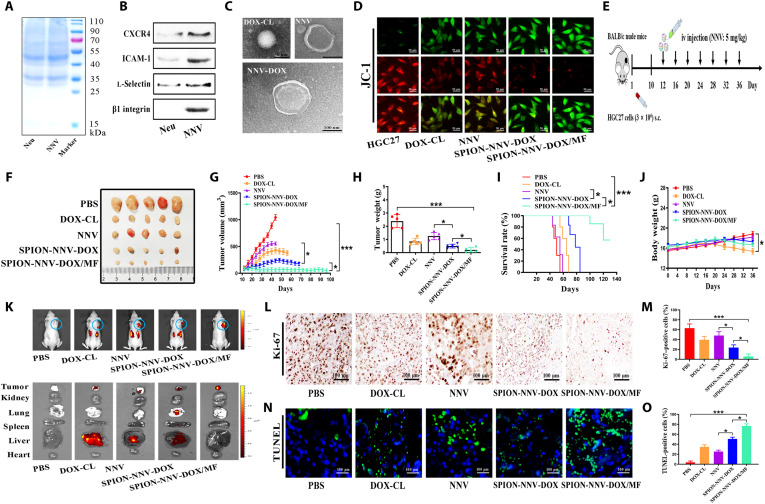
Fig. 6. SPION-modified, N-Ex–like NVs deliver DOX to inhibit tumor growth.
(A) Protein profiles of neutrophils and its derived NNV were determined by SDS–polyacrylamide gel electrophoresis. (B) Western blot analyses of neutrophil membrane–specific proteins including CXCR4, ICAM-1, l-selectin, and β1 integrin in the NNV. (C) Neutrophils incubated with DOX-CL (50 μg/ml) were collected to prepare for NVs that encapsulated DOX (NNV-DOX). The morphology of DOX-CL, NNV, and NNV-DOX were examined by TEM. Scale bar, 100 nm. (D) The effects of DOX-CL (50 μg/ml), NNV (40 μg/ml), and SPION-NNV-DOX (40 μg/ml) with or without MF on the mitochondrial membrane potential of HGC27 cells at 24 hours after the treatment were detected by JC-1 staining. (E) Schematic design for SPION-NNV-DOX treatment in subcutaneous HGC27 xenograft tumor model in BALB/c nude mice. iv, intravenous; sc, subcutaneous. (F and G) Images and sizes of subcutaneous xenograft tumors in mice treated with PBS, DOX-CL (5 mg/kg of body weight), NNV (5 mg/kg of body weight), and SPION-NNV-DOX (5 mg/kg of body weight, DOX) with or without MF under a 24-day treatment regimen (n = 5 per group). (H) Tumor weights of mice that received different treatments as indicated. (I) Survival rates of mice that received different treatments as indicated. (J) Body weights of mice in each group at the end of the experiment. (K) Represent images of ex vivo distribution of DiR-labeled SPION-NNV-DOX/MF (5 mg/kg of body weight) in major organs and tumors of mice at 3 days after the last injection. (L and M) Ki-67 staining of tumors from mice that received different treatments. Scale bars, 100 μm. (N and O) TUNEL staining for tumors from mice in different groups. Scale bars, 100 μm. One-way ANOVA for multiple groups were applied for statistical analysis. *P < 0.05 and ***P < 0.001.

