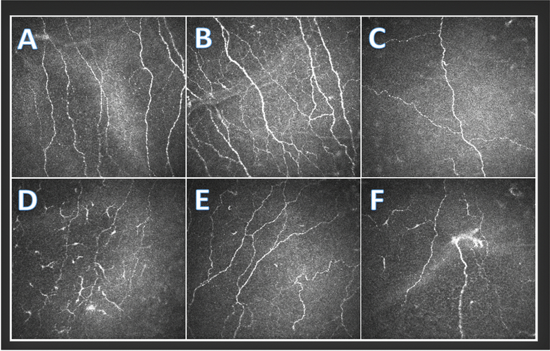
Figure 1.
In Vivo Confocal Microscopy of Sub-basal Nerves (Heidel Retina Tomograph/Cornea Rostock Module by Heidelberg Engineering, Heidelberg, Germany) depicting A) a normal nerve pattern with no dendritic cells, B) increased nerve branching, C) decreased nerve density, D) decreased nerve density and many activated dendritic cells, E) increased nerve tortuosity, and F) decreased nerve density, a probable microneuroma, and a few activated dendritic cells.

